Microscopic Examination of Urinary Sediment - Crystals, Crystals in urinary, Graff's Textbook of Urinalysis and Body Fluids
Read more: [Haematology] Microscopic Examination of Urinary Sediment - Cells
CRYSTALS
Crystals are usually not found in freshly voided urine but appear after the urine stands for a while. When the urine is supersaturated with a particular crystalline compound, or when the solubility properties of that compound are altered, the result is crystal formation. In some cases, this precipitation occurs in the kidney or urinary tract and can result in the formation of urinary calculi (stones).
Table 5-1 Properties of Crystalline Compounds
Many of the crystals that are found in the urine have little clinical significance, except in cases of metabolic disorders, calculus formation, and the regulation of medication.
The most important crystals that may be present are cystine, tyrosine, leucine, cholesterol, and sulfa. Crystals can be identified by their appearance, pH dependency, and if necessary, by their solubility characteristics (refer to Table 5-1).
Microscopic evaluation of urine is important for detection of crystals, because no chemical test detects the presence ofcrystals.
ACIDIC URINE
Those crystals which are frequently found in acidic urine are uric acid, calcium oxalate, and amorphous urates (Fig. 5-11). Crystals which occur less frequently include
Figure 5-11. Crystals frequently found in acidic urine.
calcium sulfate, sodium urates, hippuric acid, cystine, leucine, tyrosine, cholesterol, and sulfa (Fig. 5-12).
Uric Acid Crystals
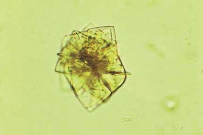
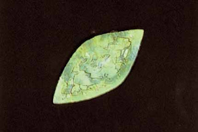
Uric acid crystals can occur in many different shapes, but the most characteristic forms are the diamond or rhombic prism (Fig. 5-13), and the rosette (Fig. 5-14 (page 66)), which consists of many crystals clustered together. They may occasionally have six sides (Fig. 5-15 (page 66)) and this form is sometimes erroneously identified as cystine. (Cystine crystals are colorless.) Uric acid crystals are usually stained with urinary pigments and are, therefore, yellow or red–brown in color. The color is frequently dependent upon the thickness of the crystal, so very thin crystals may be colorless.
Under polarized light, uric acid crystals will take on a variety of colors. The polarized crystal in Figure 5-16 (page 67) also demonstrates the layered effect that manyuric acid crystals manifest. These crystals are soluble in sodium hydroxide and insoluble in alcohol, hydrochloric acid, and acetic acid.
The presence of uric acid crystals in the urine can be a normal occurrence. It does not necessarily indicate a pathologic condition, nor does it mean that the uric acid content of the urine is definitely increased.21 Pathologic conditions in which uric acid crystals are found in the urine include gout, high purine metabolism, acute febrile conditions, chronic nephritis, and Lesch-Nyhan syndrome.
Calcium Oxalate Crystals
Calcium oxalate crystals are colorless octahedral or “envelope”-shaped crystals which look like small squares crossed by intersecting diagonal lines (Fig. 5-17). Rarely,
Figure 5-12. Other crystals found in acidic urine.
they also appear as oval spheres or biconcave disks whichhave a dumbbell shape when viewed from the side. These crystals can vary in size so that at times they are only barely discernible under high power magnification. When focusing on the typical calcium oxalate crystal, the viewer will see the “X” of the crystal popping out of the field (Fig. 5-18). Calcium oxalate crystals are frequently found in acidic and neutral urine, and occasionally they are also found in alkaline urine. They are soluble in hydrochloric acid but insoluble in acetic acid.
Calcium oxalate crystals can be present normally in the urine especially after the ingestion of various oxalate-rich food such as tomatoes, spinach, rhubarb, garlic, oranges, and asparagus. Increased amounts of calcium oxalates, particularly if they are present in freshly voided urine, suggest the possibility of oxalate calculi. Other pathologic conditions in which calcium oxalates can be present in increased numbers include ethylene glycol poisoning, diabetes mellitus, liver disease, and severe chronic renal disease.
Calcium oxalate crystals may be present in the urine following the intake of large doses of vitamin C. Oxalic acid is one of the breakdown products of ascorbic acid, and oxalic acid precipitates ionized calcium. This precipitation may also result in a decrease in the level of serum calcium.
Amorphous Urates
Urate salts of sodium, potassium, magnesium, and calcium are frequently present in the urine in a noncrystalline, amorphous form. These amorphous urates have a yellow–red granular appearance (Fig. 5-19) and are soluble in alkali and at 60 C. Amorphous urates have no clinical significance.
Figure 5-13. Uric acid crystals. Diamond or rhombic prism form (500x)
Figure 5-14. Uric acid crystals in rosette formation(500x)
Figure 5-15. Six-sided uric acid crystal (400x).
Figure 5-16. Polarized uric acid crystal. Note the layered or laminated surface (400x)
Figure 5-17. Calcium oxalate crystals (400x).
Figure 5-18. Calcium oxalate crystals. The “X” of each crystal is very prominent (500x).
Hippuric Acid Crystals
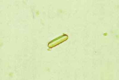
Hippuric acid crystals are yellow–brown or colorless elongated prisms or plates (Fig. 5-20). They may be so thin as to resemble needles, and they often cluster together. Hippuric acid crystals are more soluble in water and ether than are uric acid crystals.21 Hippuric acid crystals are rarely seen in the urine and have practically no clinical significance.
Sodium Urates
Sodium urates may be present as amorphous or crystalline forms (Fig. 5-21). Sodium urate crystals are colorless or yellowish needles or slender prisms occurring in sheaves or clusters. They are soluble at 60 C and only slightly soluble in acetic acid. Sodium urates have no clinical significance.
Calcium Sulfate Crystals
Calcium sulfate crystals are long, thin, colorless needles or prisms that are identical in appearance to calcium phosphate. The pH of the urine helps differentiate these two crystals, because calcium sulfate is found in acidic urine, whereas calcium phosphate is usually found in alkaline urine. Calcium sulfate is also extremely soluble in acetic acid. Calcium sulfate crystals are rarely seen in the urine and they have no clinical significance.
Cystine Crystals
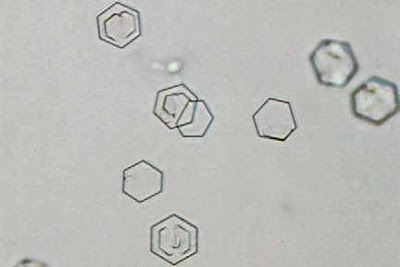
Cystine crystals are colorless, refractile, hexagonal plates with equal or unequal sides (Fig. 5-22). They may appear singly, on top of each other, or in clusters. Cystine crystals frequently have a layered or laminated appearance (Fig. 5-23). Cystine crystals are insoluble in acetic acid, alcohol, acetone, ether, and boiling water. They are soluble in hydrochloric acid and alkali, especially ammonia. Solubility in ammonia helps differentiate cystine from colorless, six-sided uric acid crystals. Cystine can be detected chemically with the sodium cyanide–sodium nitroprusside test.
The presence of cystine crystals in the urine is always important. They occur in patients with either congenital cystinosis or congenital cystinuria. Cystine crystals can form calculi.
Leucine
Leucine crystals are oily, highly retractile, yellow or brown spheroids with radial and concentric striations (Fig. 5-24). These spheroids are probably not pure leucine, because pure leucine crystallizes out as plates. Leucine is soluble in hot acetic acid, hot alcohol, and in alkali, but Leucine crystals are clinically very significant. They are found in the urine of patients with maple syrup urine disease, oasthouse urine disease,22 and in serious liver disease such as terminal cirrhosis of the liver, severe viral hepatitis, and acute yellow atrophy of the liver. Leucine and tyrosine crystals are frequently present together in the urine ofpatients with liver disease.
Tyrosine
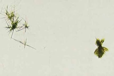
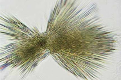
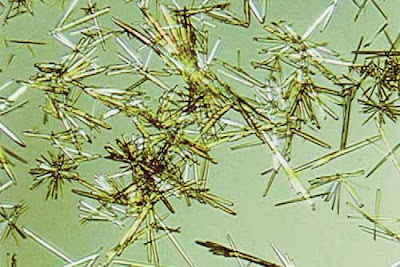
Tyrosine crystals are very fine, highly retractile needles occurring in sheaves or clusters (Figs. 5-25 and 5-26 . The needle clusters often appear to be black, especially in the center, but they may take on a yellow color in the presence of bilirubin. Tyrosine crystals are soluble in ammonium hydroxide and hydrochloric acid but insoluble in acetic acid. Tyrosine crystals can be seen in tyrosinosis and oasthouse urine disease.
Cholesterol
Cholesterol crystals are large, flat, transparent plates with notched corners (Fig. 5-27). Under polarized light they exhibit a variety of colors.24 They are soluble in chloroform, ether, and hot alcohol. At times, cholesterol crystals are found as a film on the surface of the urine instead of in the sediment.
The presence of cholesterol plates in the urine indicates excessive tissue breakdown and these crystals are seen in nephritis and nephritic conditions. They may also be present in chyluria which is the result of either thoracic or abdominal obstruction to lymph drainage, thereby causing rupture of the lymphatic vessels into the renal pelvis or urinary tract. Some of the causes of obstruction to the lymphatic flow include tumors, gross enlargement of the abdominal lymph nodes, and filariasis.
Sulfamide Drug Crystals
When sulfonamide drugs were first introduced, there were many problems with renal damage resulting from the precipitation of the drug. The newer sulfa drugs are much more soluble, even in an acid environment, and so now they rarely form crystals in the urine.
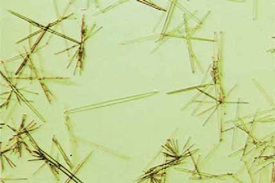
Most of the sulfonamide drugs precipitate out as sheaves of needles, usually with eccentric binding, and they may be clear or brown in color (Fig. 5-28). Two steps should be followed to confirm the presence of sulfa crystals. First, if possible, contact the nursing station (if the urine is from an inpatient) to verify that the patient is taking sulfa medication. Second, perform the lignin test for sulfonamides, which is discussed in Appendix B. Sulfoamide crystals are soluble in acetone
Radiographic Dye Crystals
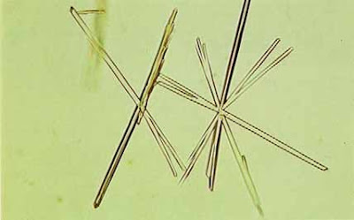
Radiographic dyes include Hypaque (Fig. 5-29) and Renografin (Fig. 5-30). Both dyes are diatrizoate meglumine plus diatrizoate sodium and can crystallize out in an acidic urine following intravenous injection for x-ray studies. Both of these dyes crystallize out as pleomorphic needles that can occur singly or in sheaves. These needles may be quite large, are often seen with brown spheres (Fig. 5-31),
and will polarize light (Fig. 5-32 (page 73)). Radiographic dyes
Figure 5-19. Amorphous urates (200x)
Figure 5-20. Hippuric acid crystal (400x).
Figure 5-21. Sodium urate crystals. These needlelike crystals are not pointed at the ends (400x)
Figure 5-22. Cystine crystal (1000x)
Figure 5-23. Cystine crystals. Several have laminated surfaces (160x)
Figure 5-24. Leucine spheroid. Note the radiating and concentric
striations as well as the brown coloring typical of these crystals (400x)
Figure 5-25. Tyrosine crystals (160x).
Figure 5-26. Same tyrosine crystals as shown in Figure 5-25, but under a higher power.
Note the fine, highly refractile needles that are typical of these crystals (1000x)
Figure 5-27. Cholesterol crystal with typical notched edges (250x).
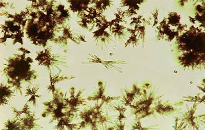
Figure 5-28. Sulfonamide crystals, yeast, and WBCs. This photograph demonstrates two typical formations of sulfa crystals: the fan or sheaf formation and sheaves with eccentric bindings (400x).
Figure 5-29. X-ray dye crystals (Hypaque) (160x).
Figure 5-30. X-ray dye crystals (Renografin) (400x).
Figure 5-31. X-ray dye crystals (Hypaque). Radiographic dyes
are fre-quently accompanied by brown spheres (160x)
are very dense, and, when present in the urine, will result in an elevated specific gravity. The presence of needle crystals in a urine with a grossly elevated specific gravity (often 1.050) is usually indicative of x-ray dye. Radiographic dyes may be present in the urine for up to 3 days following injection.
Miscellaneous Crystals
The administration of large parenteral doses of ampicillin can result in the drug precipitating out as masses of long, thin, colorless needles in acidic urine. Other drugs can occasionally result in the formation of crystals if administeredin very large doses.
In some cases of bilirubinuria, the bilirubin may crystallize out in acidic urine as red or reddish-brown needles or granules (Fig. 5-33). Bilirubin crystals are readily soluble in chloroform, acetone, acid, and alkali but are insoluble in alcohol and ether. These crystals are of no more significance than the fact that bilirubin is present in the urine.
Figure 5-32. Polarized x-ray dye crystals (160x)
Figure 5-33. Bilirubin crystals(500x).
ALKALINE URINE
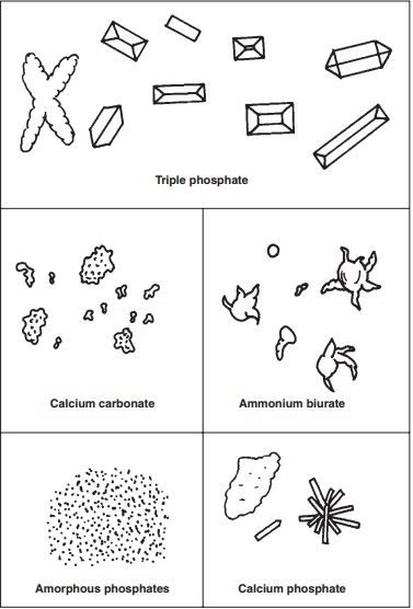
Those crystals which can be found in alkaline urine include triple phosphates (ammonium magnesium phosphates), amorphous phosphates, calcium carbonate, calcium phosphate, and ammonium biurates, also called ammonium urates (Fig. 5-34).
Triple Phosphates
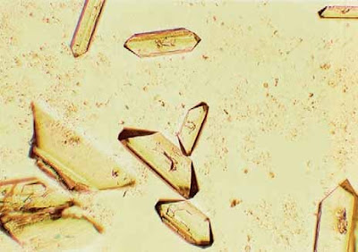
Triple phosphate (ammonium magnesium phosphate) crystals can be present in neutral and alkaline urines. Triple phosphate crystals are colorless prisms with from three to six sides that frequently have oblique ends (Fig. 5-35). Triple phosphates may sometimes precipitate as feathery or fernlike crystals. Triple phosphate crystals are soluble in acetic acid.
Triple phosphate crystals are frequently found in normal urine but can also form urinary calculi. Pathologic conditions in which they may be found include chronic pyelitis, chronic cystitis, enlarged prostate, and when the urine is retained in the bladder.
Amorphous Phosphates
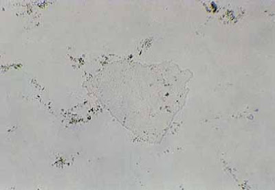
Phosphate salts are frequently present in the urine in a noncrystalline, amorphous form (Fig. 5-36). These granular particles have no definite shape and they are usually visibly indistinguishable from amorphous urates. The pH of the urine helps distinguish between these two amorphous deposits as well as does their solubility properties. Amorphous phosphates are soluble in acetic acid, whereas amorphous urates are insoluble. Amorphous phosphates have no clinical significance.
Calcium Carbonate
Calcium carbonate crystals are small, colorless crystals appearing in dumbbell or spherical forms, or in large granular masses (Fig. 5-37). Calcium carbonate crystals are larger than amorphous and, when in clumps, they may appear to have a dark color. The mass of calcium carbonate crystals, as opposed to a clump of amorphous phosphates, will also be connected together around the edges.
Calcium carbonate crystals have no clinical significance, and they will dissolve in acetic acid with the resulting evolution of carbon dioxide gas.
Calcium Phosphate
Calcium phosphate crystals are long, thin, colorless prisms and can have one pointed end, be arranged as rosettes or stars (stellar phosphates), or appear as needles (Fig. 5-38). Calcium phosphate crystals may also form large, thin, irregular plates that may float on the surface of the urine (Fig. 5-39). Calcium phosphate crystals are soluble in dilute acetic acid. These crystals may be present in normal urine, but they may also form calculi.
Ammonium Biurates
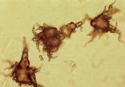
Ammonium biurate crystals, also referred to as ammonium urates, are found in alkaline and neutral urine. However, they may occasionally be found in acidic urine. Ammonium biurates are yellow–brown spherical bodies with long, irregular spicules (Fig. 5-40). Their appearance is often described with the term “thorn apple.” Ammonium biurates may also occur as yellow–brown spheroids without spicules (Fig. 5-41 (page 77)), although this form is not that common
Figure 5-34. Crystals found in alkaline urine
Figure 5-35. Triple phosphate crystals. Note the oblique ends of the prisms (200x).
Figure 5-36. Amorphous phosphates (400x)
Figure 5-37. Calcium carbonate crystals. The small arrow points out the typical “dumbbell”
form which is next to a large mass of calcium carbonate crystals
Figure 5-38. Calcium phosphate crystals (400x)
Figure 5-39. Calcium phosphate plate or phosphate sheath (200x)
Figure 5-40. Ammonium biurate crystals (500x).
Figure 5-41. Ammonium biurate crystals without spicules(500x).
Ammonium biurates dissolve by warming and are soluble in acetic acid, with the formation of colorless uric acid crystals after standing. The addition of sodium hydroxide will liberate ammonia. Ammonium biurates are abnormal only if found in freshly voided urine.
REFERENCES
Lillian A. Mundt and Kristy Shanahan, Graff's Textbook of Urinalysis and Body Fluids, Second Edition 2011















COMMENTS