
Atlas of Otitis media - Acute otitis and otitis media with effusion, Otitis media, Acute otitis and otitis media with effusion, Cholesteatoma, atlas in medical, tuyenlab.net
 |
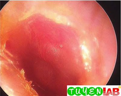
| FIGURE 1. Acute otitis media in the left ear of a 15-month-old patient with marked erythema and bulging of the tympanic membrane. The malleus and light re ex are not visible. |
 in the right ear") |
| FIGURE 2. Otitis media with effusion (OME) in the right ear. Note multiple airuid levels in this slightly retracted, translucent, nonerythematous tympanic membrane. |
 and straightening of the handle of the malleus as the retraction pulls the bone upward") |
| FIGURE 3. Otitis media with effusion in the left ear showing retraction of the tympanic membrane (TM) and straightening of the handle of the malleus as the retraction pulls the bone upward. |
 |
| FIGURE 4. Early acute otitis media at the stage of eustachian tube obstruction. Note the slight retraction of the tympanic membrane (TM), the more horizontal position of the malleus, and the prominence of the lateral process. |
 |
| FIGURE 5. Acute otitis media, stage of suppuration. Note presence of purulent exudate behind the tympanic membrane (TM), the outward bulging of the TM, prominence of the posterosuperior portion of the drum, and generalized TM edema. The white area is tympanosclerosis from a previous infection. |

 |
| FIGURE 6. A. Normal right tympanic membrane with comparison using. B. Normal bony landmarks of the inner ear. The ossicles were removed in this dissection. |
 |
| FIGURE 7. Cholesteatoma. |
 |
| FIGURE 8. Primary acquired cholesteatoma with debris removed from the attic retraction pocket. |
 |
| FIGURE 22-9 Bullous myringitis can be differentiated from otitis media with effusion by identifying serouslled bulla on the surface of the tympanic membrane (TM) |

 |
| FIGURE 10. A. Mastoiditis in a young boy with recurrent otitis media. Note the erythema and swelling behind the ear. The ear is sticking out more than the other side. B. Surgical drainage was performed. |
 |
| FIGURE 11. Traumatic perforation of the left tympanic membrane. |
 of a 9-year-old girl with recurrent acute otitis media and chronic TM retractions prior to polyethylene (PE) tube placement")
 tube is placed in the anterior-inferior quadrant of the TM of a 9-yearold girl with recurrent acute otitis media") |
| FIGURE 12. A. Left tympanic membrane (TM) of a 9-year-old girl with recurrent acute otitis media and chronic TM retractions prior to polyethylene (PE) tube placement. The circular area near the center of the TM is caused by the TM being retracted against the promontory of the medial wall of the middle ear. B. A fluoroplastic polyethylene (PE) tube is placed in the anterior-inferior quadrant of the TM of a 9-yearold girl with recurrent acute otitis media. It is black because it is impregnated with silver oxide to retard the growth of bacterial microfilms. |
 |
| FIGURE 13. Tympanosclerosis as the result of previous recurrent episodes of otitis media and polyethylene (PE) tube placement. |
This is only a part of the book : Color Atlas of Pediatrics 1st Edition of authors: Richard P. Usatine, MD; Camille Sabella, MD; Mindy Ann Smith, MD; E.J. Mayeaux, Jr., MD; Heidi S. Chumley, MD and Elumalai Appachi, MD, MRCP (UK). If you want to view the full content of the book and support author. Please buy it here: https://goo.gl/BEp0yD


COMMENTS