Atlas of congenital anomalies of the head and neck, congenital anomalies of the head and neck, Pediatric, ATLAS IN MEDICAL, TUYENLAB.NET
 |
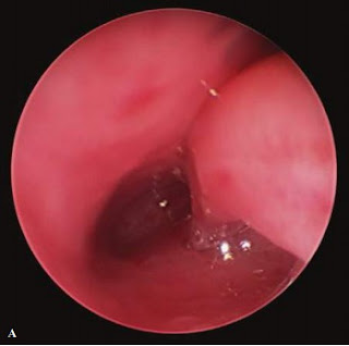
| Fig 3. Left unilateral choanal atresia seen on endoscopy (A, B). Note inferior turbinate laterally (A). The right side is unaffected (C) with a patent choanal opening seen behind the turbinate. |
 |
| Fig 4. Predominantly nasal dermoid in an infant causing unilateral nasal obstruction. No intracranial extension was noted. Note the small dimple on nasal tip. |
 |
| Fig 5. Microtia (underdeveloped pinna) in a child. |
 |
| Fig 7. 3 week old infrant with severe micrognathia and upper airway obstruction, prior to mandibular distraction, on preoperative, pro le view (A) and on CT scan with 3D reconstruction (B). |
 |
| Fig 8. Micrognathia in an infant, which ultimately required mandibular distraction for persistent respiratory distress and poor growth. |
 |
| Fig 10. Congenital epulis (benign tumor on the gingival or alveolar mucosa) arising from maxillary alveolar ridge in a newborn, on lateral (A) and primarily frontal (B) views. |
 |
| Fig 11. Oral dermoid cyst in a 2-year-old child being surgically excised from the oor of the mouth. |
 |
| Fig 12. Naso-oropharyngeal soft tissue stenosis in a young child with Mobius syndrome. Endoscopic view with exible scope shows narrowed caliber, normal larynx in the distance. |
 |
| Fig 14. Thyroglossal duct cyst in a 2-year-old girl, as seen on frontal (A) and lateral (B) views. |
 |
| Fig 15. Acutely infected thyroglossal duct cyst in a teenager. This requied incision and drainage prior to definitive Sistrunk procedure. (excision of cyst with tract and middle third of hyoid bone). |
 |
| Fig 16. Infected right preauricular sinus with abscess in a young girl. |
 |
| Fig 17. First Branchial Anomaly. Intraoperative view of Type 1 first branchial anomaly with duplicated cartilage and sinus. This was excised along with small cyst and external canal reconstructed. |
 |
| Fig 19. Cleft palate as seen intraoperatively prior to repair (A) and immediately after repair (B). |
 |
| Fig 20. Bilateral cleft lip with prominent primaxilla, prior to repair (A), and 1 week after repair (B), |
This is only a part of the book : Color Atlas of Pediatrics 1st Edition of authors: Richard P. Usatine, MD; Camille Sabella, MD; Mindy Ann Smith, MD; E.J. Mayeaux, Jr., MD; Heidi S. Chumley, MD and Elumalai Appachi, MD, MRCP (UK). If you want to view the full content of the book and support author. Please buy it here: https://goo.gl/BEp0yD


COMMENTS