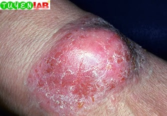
These are pictures of Contact Dermatitis and Drug Eruptions (part 1). This is a part in Contact Dermatitis and Drug Eruptions of the Skin Clinical Atlas 1st Edition 2018 book
Contact dermatitis and cutaneous drug eruptions are both skin conditions triggered by external agents that are either in direct contact with the skin or ingested. Contact dermatitis shares many features with atopic dermatitis and eczema, including pruritus, erythema, scaling, oozing, and crusting.
However, in the case of contact dermatitis, major clues to the diagnosis are the shapes found on the skin that demonstrate the particular locations of contact by the external irritant or allergic stimuli. These shapes can be classically linear like Toxicodendron dermatitis (poison ivy); geometric such as seen secondary to adhesives; in a splattered pattern secondary to liquid irritants (e.g., acids); or in the case of airborne triggers such as fragrances, concentrated on the face, neck, and other exposed regions of the body. Noting the location of the affected body part or parts is especially helpful in diagnosing contact dermatitis, such as the lower abdomen and ears in nickel dermatitis, dorsal feet in shoe dermatitis, and eyelids in dermatitis secondary to ingredients in nail polish. Other contact reactions can be more widespread, as is the case in some patients with atopic dermatitis and concomitant contact dermatitis due to preservatives or fragrances in lotions and detergents, respectively.
Cutaneous drug eruptions can be quite varied in their presentations, ranging from the sheets of micropustules overlying patches of broad erythema seen in acute generalized exanthematous pustulosis to the hemorrhagic mucosal erosions and vesicobullous lesions seen in Stevens-Johnson syndrome and toxic epidermal necrolysis. It is imperative to screen patients for associated features on physical examination such as fever, lymphadenopathy, and facial edema that can help distinguish a systemic drug hypersensitivity reaction such as drug reaction with eosinophilia and systemic symptoms (DRESS) from a simple morbilliform drug eruption. Blood work may also be needed in some patients with suspected DRESS to screen for eosinophilia, atypical lymphocytes, and evidence of end organ damage. Other drug eruptions may be more unique, such as the striking, deeply erythematous, circular patches and plaques of fxed drug eruptions or painful palmoplantar plaques of toxic erythema of chemotherapy. Because new medications are being produced every year, it is also important to remain knowledgeable about the diverse idiosyncratic drug eruptions such as the erosive psoriasiform scalp and posterior auricular plaques seen in individuals treated with tumor necrosis factor inhibitors.
This portion of the atlas will highlight the cutaneous patterns created by contact dermatitis, as well as the variety of skin fndings seen in the setting of both mild and severe drug eruptions.
Fig. 6.1 Cement burns.
Fig. 6.2 Acid burn.
Fig. 6.3 Drip burn from acid solution.
Fig. 6.4 Senna burn
Fig. 6.5 Pesticide burn
Fig. 6.6 Irritant dermatitis from kerosene
Fig. 6.7 Positive patch test.
Fig. 6.8 Poison ivy dermatitis; note linear lesions.
Fig. 6.9 Poison ivy dermatitis; note linear lesions
Fig. 6.10 Poison ivy dermatitis
Fig. 6.11 Poison ivy dermatitis.
Fig. 6.12 Poison ivy dermatitis
Fig. 6.13 Tea tree oil dermatitis.
Fig. 6.14 Clothing contact dermatitis
Fig. 6.15 (A) Blue dye dermatitis. (B) Resolved when using diapers without blue dye
Fig. 6.16 Acute shoe dermatitis
Fig. 6.17 Chronic shoe dermatitis
Fig. 6.18 (A) Nickel dermatitis. (B) With earrings on.
Fig. 6.19 Nickel dermatitis.
Fig. 6.20 Black dermatographism
Fig. 6.21 Oral lichenoid dermatitis due to gold
Fig. 6.22 Stomatitis due to dental impression material allergy
Fig. 6.23 (A) Rubber dermatitis. (B) Culprit swim goggles.
Fig. 6.24 Earbud dermatitis.
Fig. 6.25 Contact allergy to adhesive in bandage
Fig. 6.26 Hand dermatitis due to aromatherapy
Fig. 6.27 Eyelid dermatitis secondary to nail polish.
Fig. 6.28 Hair dye dermatitis.
Fig. 6.29 Vulvar dermatitis secondary to preservatives
Fig. 6.30 Clonidine patch allergy
Fig. 6.31 Allergy to benzocaine
Fig. 6.32 Allergy to topical antibiotic
Fig. 6.33 Leg ulcer with contact dermatitis due to topical antibiotic allergy
Fig. 6.34 Contact allergy to topical steroids
Fig. 6.35 Contact cheilitis to Blistik.
Continue... -> Read more:
Atlas of Contact Dermatitis and Drug Eruptions (part 2)
Atlas of Contact Dermatitis and Drug Eruptions (part 3)
REFERENCES
Andrews' Diseases of the Skin Clinical Atlas 1st Edition 2018
Atlas of Contact Dermatitis and Drug Eruptions (part 3)
REFERENCES
Andrews' Diseases of the Skin Clinical Atlas 1st Edition 2018


COMMENTS